Discovery of a Two-Layered Cyst Wall Structure Responsible for the Transmission of Amoebic Dysentery
A collaborative research group led by Assistant Professor Tam Kha Vo and Professor Fumika Mi-ichi of the Institute of Tropical Medicine (NEKKEN), Nagasaki University, together with Professor Hiroki Yoshida of the Faculty of Medicine, Saga University, has elucidated the formation mechanism of the cyst wall that surrounds the infectious cyst responsible for the transmission of Entamoeba histolytica, the causative agent of amoebic dysentery—a parasitic infection that can also occur in Japan.
Their analysis revealed that the cyst wall, previously believed to consist of a single layer composed of chitin fibers and structural proteins, actually has a two-layered structure. The newly identified inner layer contains hydrophobic components, likely lipids, which may play an important role in high environmental resistance and barrier function of Entamoeba cysts (Fig. 1).
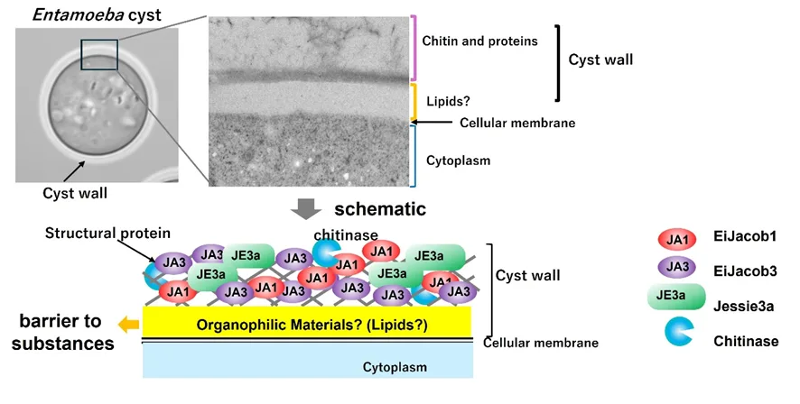
Figure 1 Overview of the Study
Amoebic dysentery is caused by the parasitic protozoan Entamoeba histolytica. It is estimated that about 50 million people worldwide are infected each year, with 40,000–70,000 deaths annually. In Japan, approximately 1,000 clinical cases are reported each year. Because treatment options are limited, associated with side effects, and no effective vaccine is currently available, the development of new therapeutic strategies is an urgent need.
The primary route of human infection is the oral ingestion of cysts. These cysts are dormant structures surrounded by a thick cyst wall that enables them to survive outside the host under harsh conditions, including dry environments and gastric acid. They are excreted in feces and can infect new hosts through contaminated water or food.
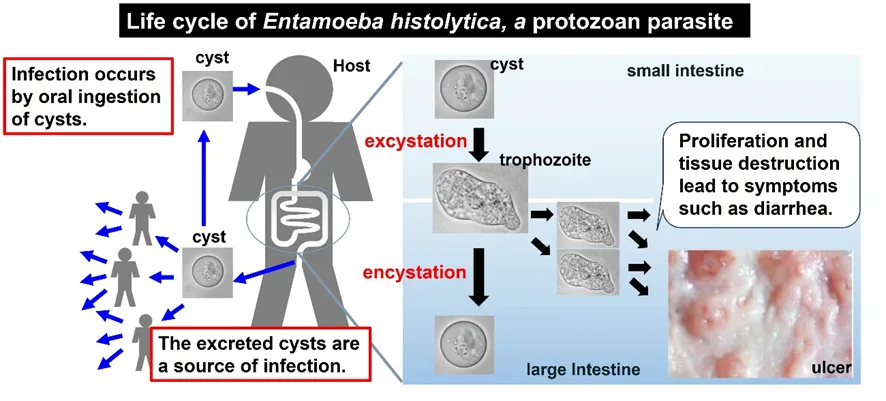
Figure 2 Life Cycle of Entamoeba histolytica
In this study, the researchers generated antibodies against the structural proteins Jacobs and Jessies, which are components of the cyst wall, and analyzed their localization using fluorescence microscopy and electron microscopy. The results showed that chitin fibers and structural proteins are initially distributed uniformly from the cell membrane outward, but gradually become concentrated near the cell surface as cyst formation progresses. In addition, a layer of hydrophobic components soluble in organic solvents was found to form between the cell membrane and the chitin layer.
Further analyses revealed that although cysts are highly resistant to physical stress, they show high sensitivity to organic solvents. This finding suggests that the hydrophobic components in the inner layer of the cyst wall are essential for maintaining cyst structure and function.
This discovery may lead to new strategies targeting the cyst wall structure. For example, removal of cysts using organic solvents such as ethanol could provide an effective approach to preventing transmission.
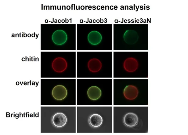
Figure 3 Localization analysis using each antibody
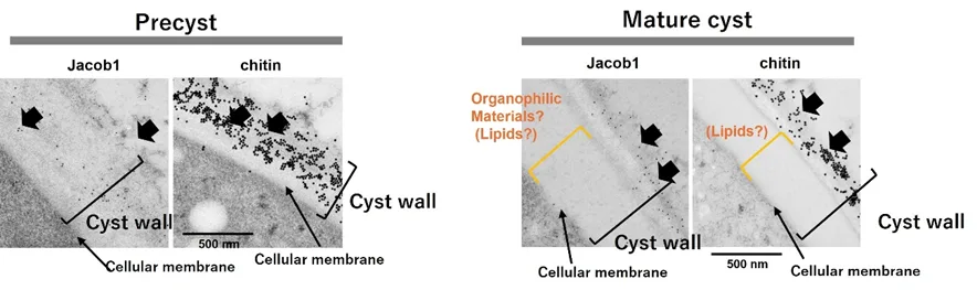
Figure 4 Localization analysis by immunoelectron microscopy
Journal: PLoS Pathogens
Title: Trafficking and organization of cyst wall components into a robust biphasic structure in Entamoeba
Authors: Tam Kha Vo, Hiroki Yoshida, Fumika Mi-ichi
DOI: 10.1371/journal.ppat.1013940
For more details, please refer to the full article published in PLoS Pathogens.
This research was supported by the Kaketsuken Research Grant “Functional Analysis of Ultra-long-chain Dihydroceramide Produced by Entamoeba histolytica in Dormancy” (Principal Investigator: Fumika Mi-ichi) and the Japan Agency for Medical Research and Development (AMED) programs, including the project “Lipid Metabolism Analysis of Entamoeba histolytica Based on Lipidomics Metadata: Providing Biochemical and Physiological Insights and Drug Targets” (2021–2024), as well as the ongoing project “Creation of an Infectious Disease Drug Discovery Platform Based on a Multidimensional Understanding of the Host–Parasite Interaction in Entamoeba histolytica Infection,” among others.


